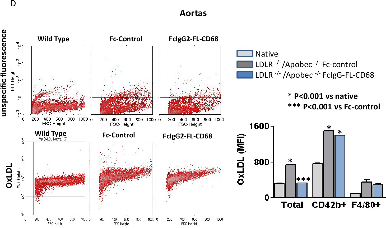
Fig. 5. oxLDL content was determined in vascular cells from the aorta from these 3 mouse groups. In representative scatter blots unspecific fluorescence of cells or oxLDL positive fluorescence of vascular cells from wild type mice, Fc-control treated and FcIgG2-FL-CD68 treated of LDLR-/-/ApoBec-/- mice are shown. Gating threshold is depicted derived from the isotype controls and applied for the specific fluorescence. oxLDL content is quantified in vascular cells from aorta homogenization (total), in cells positive for the platelet marker CD42 and the macrophage marker F4/80 measured by FACS with anti oxLDL antibodies. The means ± SEM of 5 animals are shown. * indicates statistical significance of p<0.001 versus native mice and *** against Fc-control treated of LDLR-/-/ApoBec-/- mice.